smooth muscle fiber labeled
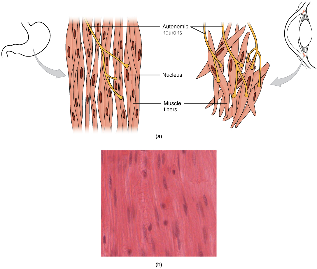
This diagram shows the structure of smooth muscle. To the left of the. 8 Images about This diagram shows the structure of smooth muscle. To the left of the : This diagram shows the structure of smooth muscle. To the left of the, Muscle fiber, Skeletal muscle, and Contraction [USMLE] - YouTube and also Muscle Fiber Tears-Sam Oster Hour 2.
This Diagram Shows The Structure Of Smooth Muscle. To The Left Of The
 oerpub.github.io
muscle smooth involuntary tissue muscles types anatomy voluntary diagram cells tissues cell labelled found physiology eye human body respiratory difference
Muscle Fiber, Skeletal Muscle, And Contraction [USMLE] - YouTube
![Muscle fiber, Skeletal muscle, and Contraction [USMLE] - YouTube](https://i.ytimg.com/vi/35f8Vzd843U/maxresdefault.jpg) www.youtube.com
www.youtube.com
muscle skeletal fiber contraction
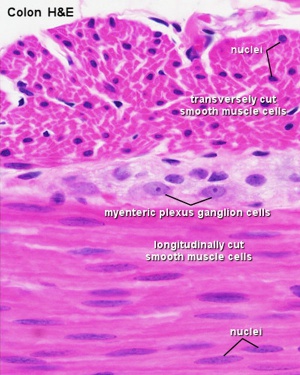
Neural Crest - Enteric Nervous System - Embryology
 embryology.med.unsw.edu.au
muscle smooth histology cells system embryology development tract tissue gastrointestinal nervous neural tissues enteric crest jejunum longitudinal file myenteric plexus
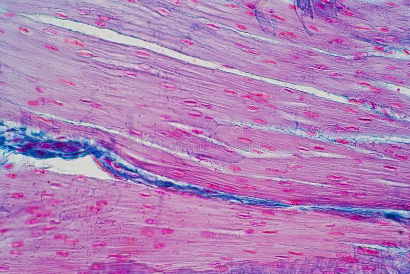
Histology Of Human Smooth Muscle Under Microscope View For Education
 www.dreamstime.com
muscle microscope under smooth human histology education
3. Skeletal Muscle
 www.bristol.ac.uk
muscle skeletal longitudinal section slide tissue microns bar striations tranverse figure pulpbits previous
Chapter 7, Page 5 - HistologyOLM 4.0
 stevegallik.org
muscle skeletal section cross histology tissue normal microscopic histologyolm stevegallik study preparation chapter
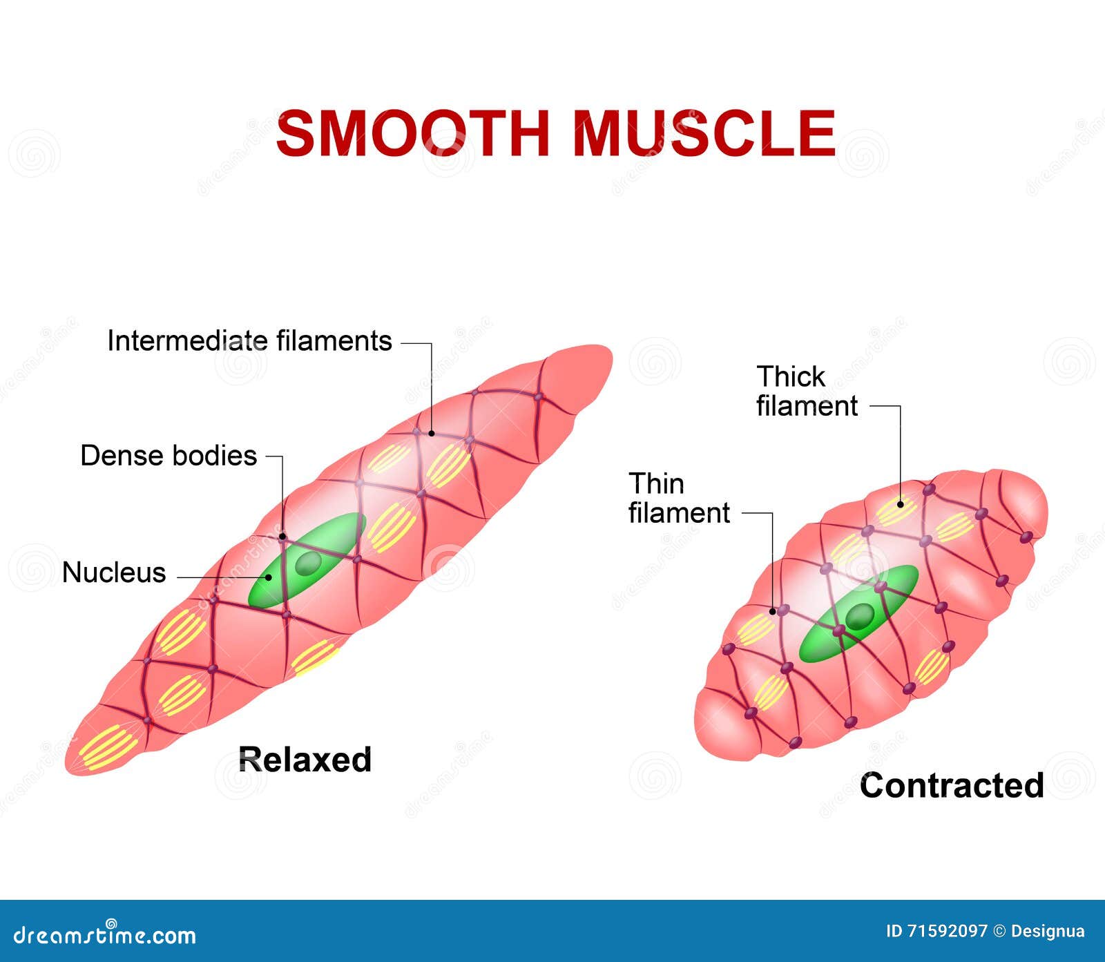
Smooth Muscle Tissue Cartoon Vector | CartoonDealer.com #71592097
 cartoondealer.com
muscolo structure liscio lisse spierweefsel muskel gewebe vlot silkespapper relaxers muskels glatten cellula tessuti skeletal slät innervation cardiaque schema vectorstock
Muscle Fiber Tears-Sam Oster Hour 2
 www.thinglink.com
micro oster
Muscle skeletal fiber contraction. Muscle fiber tears-sam oster hour 2. Micro oster