skeletal muscle fiber histology
Skeletal Muscle Fibers Cross Section Shows Peripheral Nuclei And. 16 Pictures about Skeletal Muscle Fibers Cross Section Shows Peripheral Nuclei And : File:Sarcomere animation.gif - Embryology, Skeletal Muscle Histology - Embryology and also Skeletal muscle tissue: Histology | Kenhub.
Skeletal Muscle Fibers Cross Section Shows Peripheral Nuclei And
 www.gettyimages.de
skeletal nuclei fibers peripheral myofibrils
Skeletal Muscle Histology - Embryology
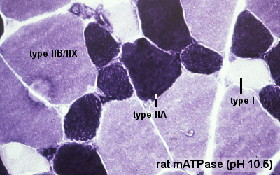 embryology.med.unsw.edu.au
muscle types fiber skeletal histology fibre embryology file development tissue em slides system unsw med edu
A Single Skeletal Muscle Fiber In Cross Section 250x High-Res Stock
 www.gettyimages.com
skeletal fiber muscle cross single
Muscle Tissue Page 1
 audilab.bmed.mcgill.ca
muscle tissue skeletal drawing cross fibres longitudinal histology paintingvalley sections right figure drawings reticular illustrated
Basic Histology -- Skeletal Muscle, Longitudinal Section
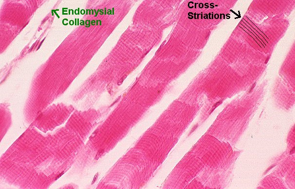 www.pathguy.com
muscle skeletal longitudinal section histology endomysium cross histo basic pronunciations states united
Skeletal Muscle Tissue: Histology | Kenhub
:watermark(/images/logo_url.png,-10,-10,0):format(jpeg)/images/anatomy_term/epimysium/pZJdNC0MZyeIzfxAXmbng_Epimysium.png) www.kenhub.com
www.kenhub.com
skeletal histology epimysium kenhub connective
Basic Histology -- Skeletal Muscle, Longitudinal Section
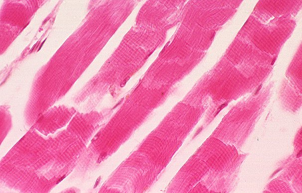 www.pathguy.com
muscle skeletal histology longitudinal section cross histo basic striations
Muscle: The Histology Guide
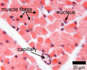 histology.leeds.ac.uk
muscle skeletal histology tissue types muscles transverse ts section cells gross anatomy shows through slides
File:Sarcomere Animation.gif - Embryology
 embryology.med.unsw.edu.au
skeletal muscle histology sarcomere tissue file cell embryology band bands sarcomeres myosin animation showing development light unsw med edu isotropic
Histology A560
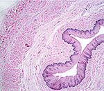 medsci.indiana.edu
a464 muscle fibers histology a560 nucleus skeletal diameter smaller than type medsci indiana edu visceral smooth
Pathology Outlines - Histology-skeletal Muscle
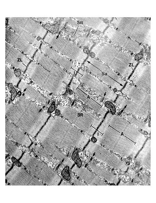 www.pathologyoutlines.com
muscle skeletal em histology contractile filament resulting arrangement fiber within band pathology
File:Cardiac Muscle EM04.jpg - Embryology
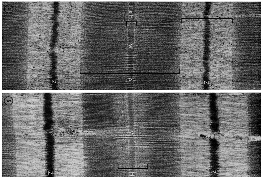 embryology.med.unsw.edu.au
muscle cardiac em04 file embryology resolutions unsw med edu
Muscle - Slide #3
 education.med.nyu.edu
muscle fibers section cross skeletal magnification unknown
Histology A560
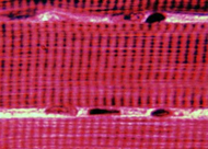 medsci.indiana.edu
muscle skeletal
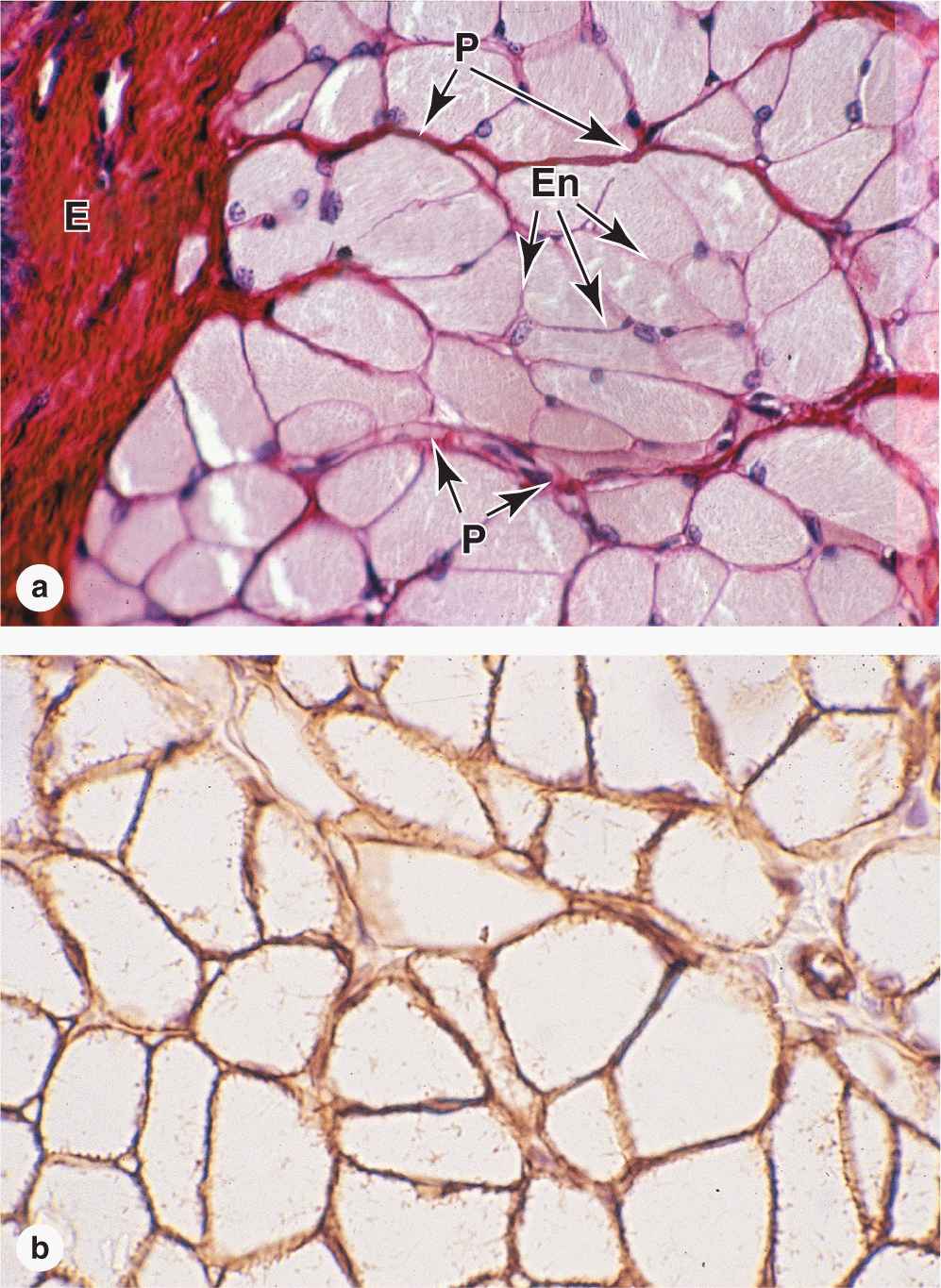
Muscle Tissue | Basicmedical Key
 basicmedicalkey.com
muscle tissue skeletal endomysium figure
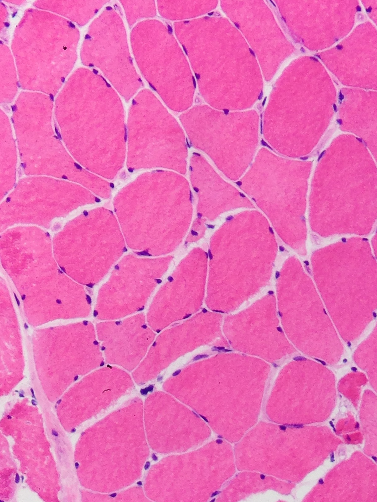
Pathology Outlines - Neurogenic Atrophy
 www.pathologyoutlines.com
atrophy skeletal neurogenic pathology biopsy outlines atrophic myofibers clumps angulated
Muscle skeletal em histology contractile filament resulting arrangement fiber within band pathology. File:cardiac muscle em04.jpg. Skeletal muscle histology sarcomere tissue file cell embryology band bands sarcomeres myosin animation showing development light unsw med edu isotropic