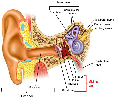
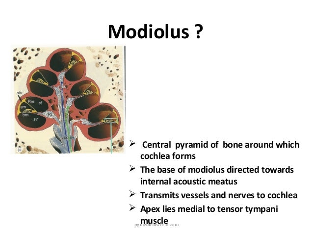
modiolus ear anatomy
18: Ear | Pocket Dentistry. 15 Images about 18: Ear | Pocket Dentistry : PPT - ANATOMY AND PHYSIOLOGY OF THE EXTERNAL EAR, MIDDLE EAR AND INNER, Human Ear Anatomy - Parts of Ear Structure, Diagram and Ear Problems and also Inner ear: Anatomy | Kenhub.
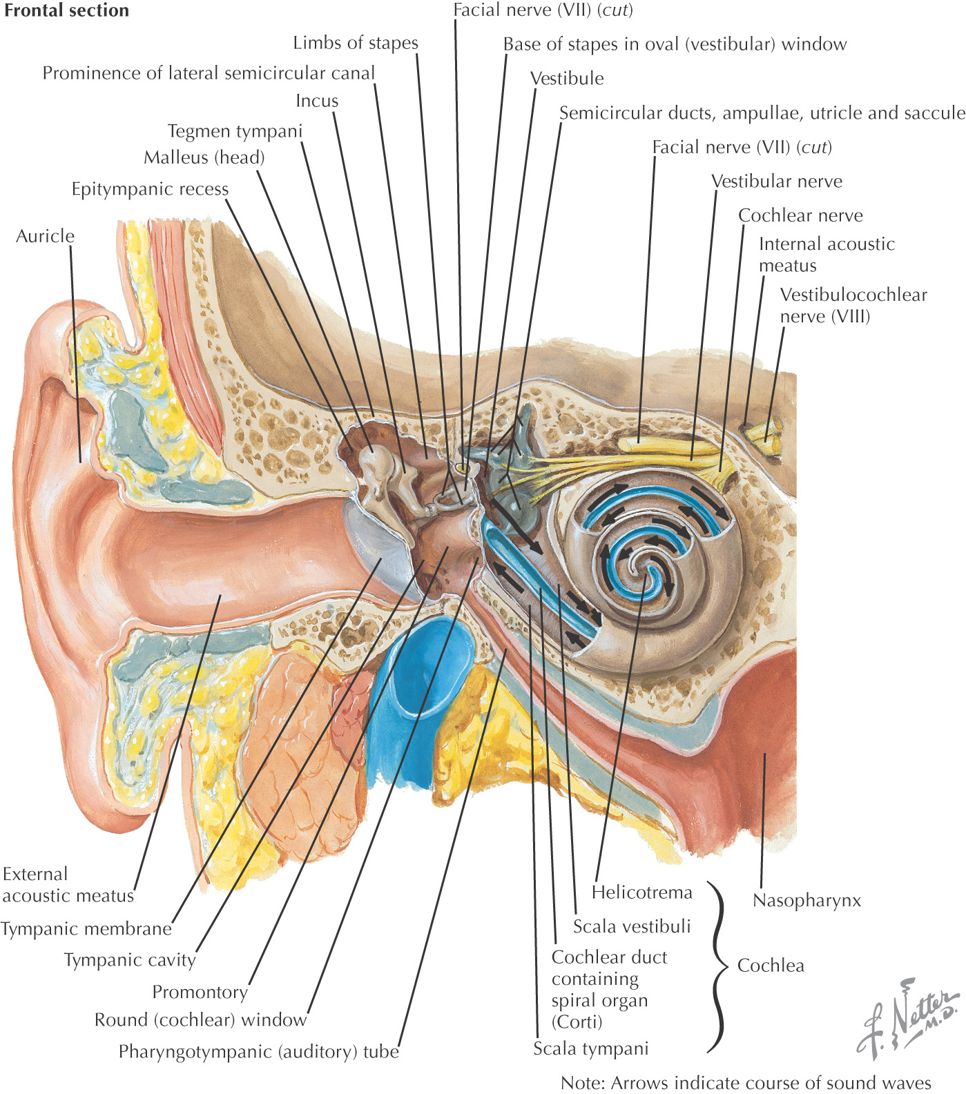
18: Ear | Pocket Dentistry
 pocketdentistry.com
ear internal meatus acoustic nerve enters vestibulocochlear via pocketdentistry
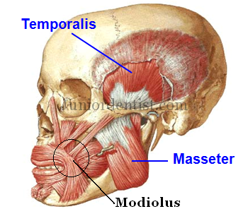
What Is Modiolus Of Face And Mouth
 www.juniordentist.com
mouth modiolus face muscles temporalis muscle facial masseter neck head origin jaw temporal insertion mandible masticatory angle dentist point process
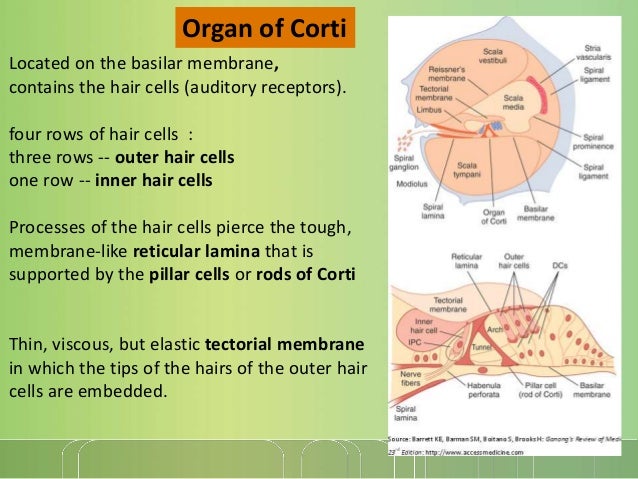
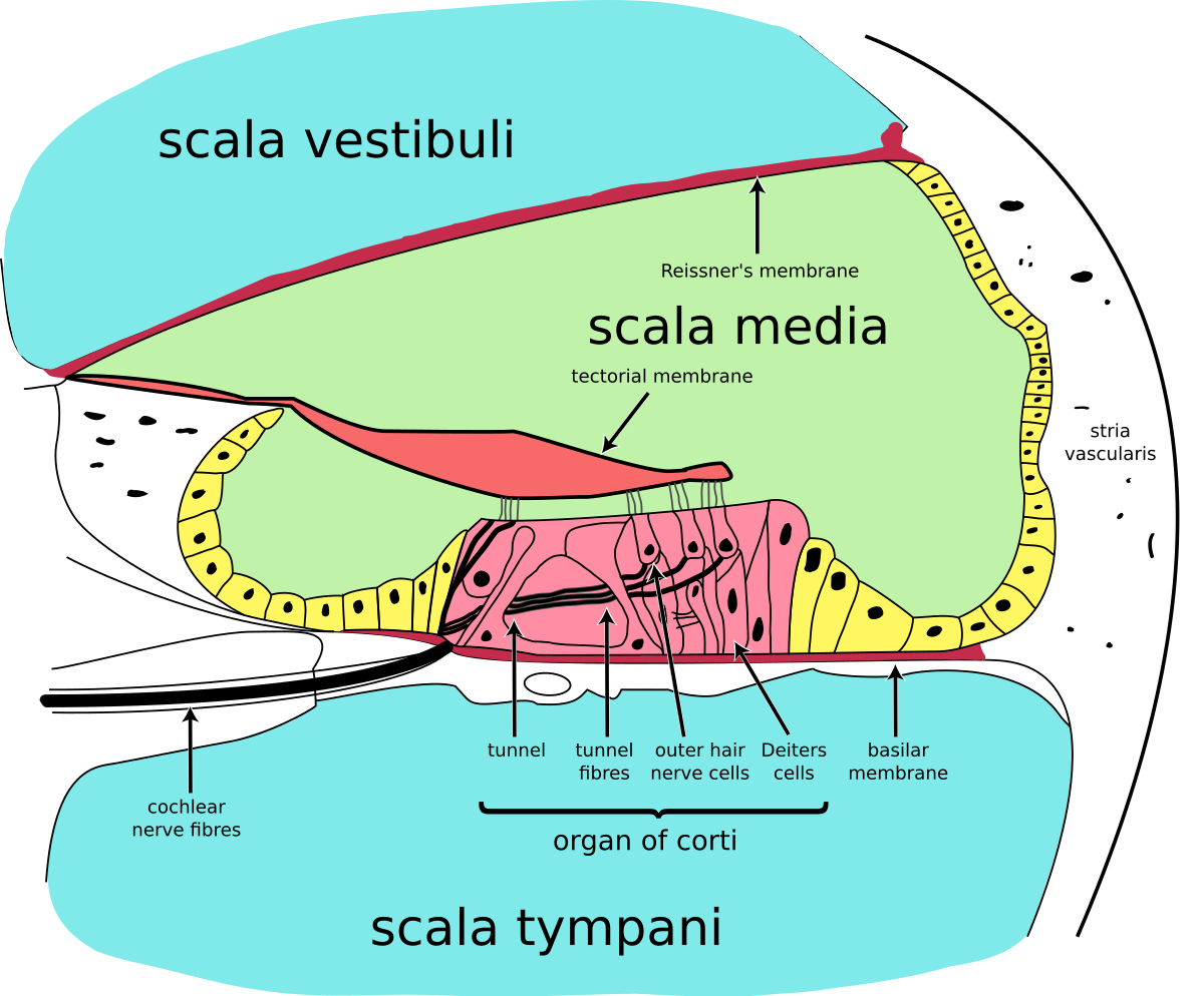
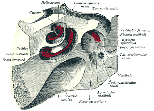
Cochlear Anatomy
 www.d.umn.edu
cochlea scala tympani vestibuli ear spiral inner cochlear anatomy lamina bony into dividing naturalphenomena science hearing membrane
Structure And Function Of Human Ear Class Eleven Biology
 www.excellup.com
ear structure function biology attached ossicles inner malleus membrane tympanic chain
Physiology Of External, Middle And Inner Ear
 www.slideshare.net
external
PPT - ANATOMY AND PHYSIOLOGY OF THE EXTERNAL EAR, MIDDLE EAR AND INNER
 www.slideserve.com
anatomy ear external cochlea ppt physiology inner middle powerpoint presentation modiolus
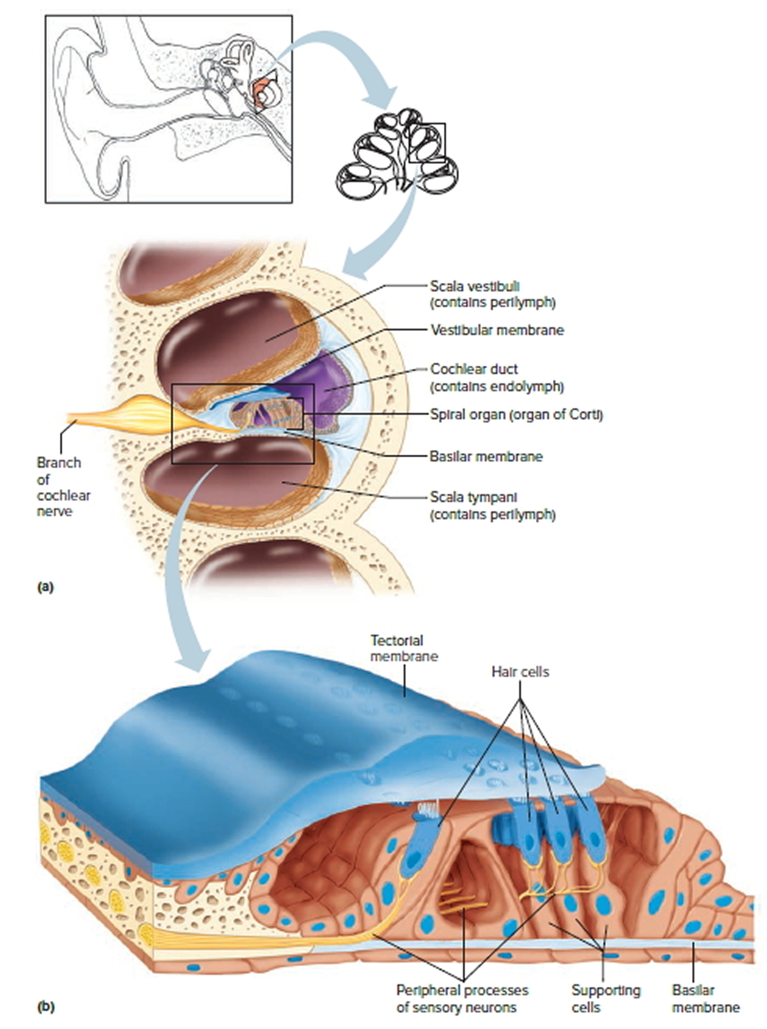
Cochlea - Wikipedia
 en.wikipedia.org
cochlea section cross membrane basilar cochlear wikipedia nerve scala ear coclea anatomy inner anatomia structures system sound nerves
Nerve Racking - Lesson - Www.teachengineering.org
 www.teachengineering.org
nerve human ear lesson parts cochlear racking auditory malleus cub drawing tube
Ear Myoclonus / Stapedial Muscle Spasm: Ear Myoclonus Info
 rockys-ear.blogspot.com
ear spasm muscle myoclonus stapedial canal tensor tympani
Anatomy Of Inner Ear
 www.slideshare.net
modiolus cochlea cochlear central
Inner Ear: Anatomy | Kenhub
cochlea ear inner cochlear kenhub pathway auditory anatomy system ventral anterior spiral oval window apex library
Anatomy Of Inner Ear
 www.slideshare.net
membranous
Ear Anatomy
 fpnotebook.com
anatomy ear tube tympanic tympani eustachian tensor cavity carotid artery middle internal petrous torus auditory portion muscle fpnotebook radiopaedia ligament
Vestibule Of The Ear - Wikidoc
 www.wikidoc.org
vestibule cochlea ear labyrinth
Human Ear Anatomy - Parts Of Ear Structure, Diagram And Ear Problems
 healthjade.com
cochlea ear human organ spiral section parts cross anatomy diagram structure note figure
Cochlea ear inner cochlear kenhub pathway auditory anatomy system ventral anterior spiral oval window apex library. Cochlea section cross membrane basilar cochlear wikipedia nerve scala ear coclea anatomy inner anatomia structures system sound nerves. Anatomy of inner ear