labeled cardiac muscle
Anatomy Of Cardiac Muscles. 16 Pictures about Anatomy Of Cardiac Muscles : Cardiac muscle (labeled) | Flickr - Photo Sharing!, Heart: Anatomy & Pathology and Components of the CVS | Lecturio and also Cardiac muscle.
Anatomy Of Cardiac Muscles
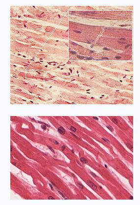
General Anatomy And Physiology Of A Human: TEAS || RegisteredNursing.org
 www.registerednursing.org
anatomy physiology human general teas cardiac muscle
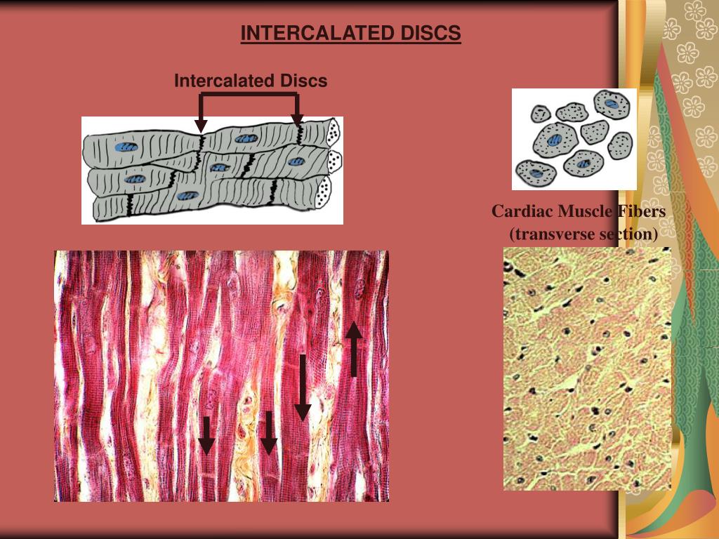
PPT - Cardiac Muscle PowerPoint Presentation, Free Download - ID:6182155
 www.slideserve.com
cardiac muscle intercalated discs ppt powerpoint presentation
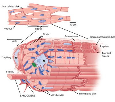
Cardiac Muscles Properties - Morphology ~ Medicine Hack
 www.medicinehack.com
cardiac otot jantung ciri morphology rangka perbedaan physiology penyusun jaringan hisham lurik sebutkan mitochondria annotated skeletal fibrils karakteristik
Heart Muscle Dr.Jastrow's Electron Microscopic Atlas
 www.uni-mainz.de
intercalated em muscle heart electron disk
Heart: Anatomy & Pathology And Components Of The CVS | Lecturio
 www.lecturio.com
cardiac muscle anatomy electrical activity heart cells cell structure tissue physiology gap junctions diagram intercalated components tubules muscles discs myofilaments
Skeletal Muscle Histology - Embryology
 embryology.med.unsw.edu.au
skeletal muscle histology tissue sarcomere cell file band bands sarcomeres myosin embryology animation showing light development isotropic unsw med edu
Chapter 7, Page 7 - HistologyOLM 4.0
 stevegallik.org
histology
HISTOLOGY OF SKELETAL MUSCLE - YouTube
 www.youtube.com
skeletal muscle histology
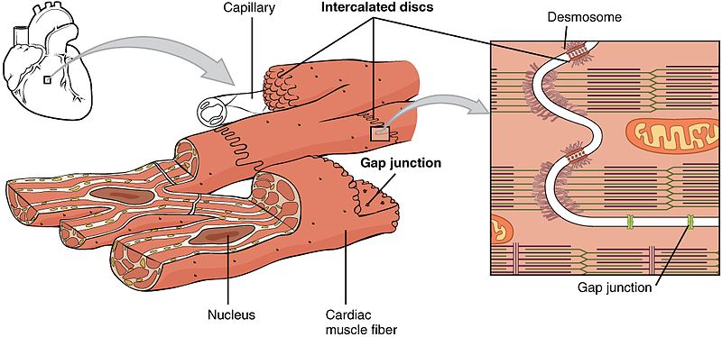
Structure Of Cardiac Muscle - Myopathy - TeachMePhysiology
 teachmephysiology.com
cardiac muscle structure diagram overall fig showing
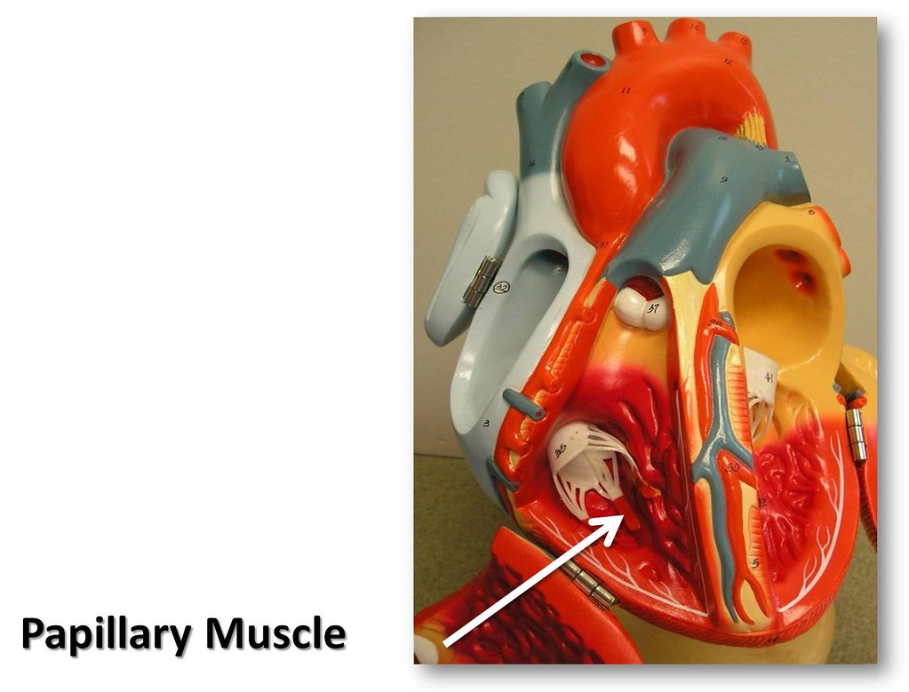
Papillary Muscle - The Anatomy Of The Heart Visual Atlas, … | Flickr
 www.flickr.com
septum heart interventricular chordae tendineae anatomy papillary muscle visual flickr atlas right weekly commons recent galleries ventricle
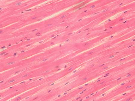
Cardiac Muscle
 www.eugraph.com
cardiac histology muscle tissues tissue lab slide cells human final photomicrographs anatomy quia physiology
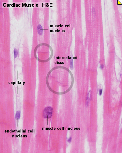
Cardiac Muscle Histology - Embryology
 embryology.med.unsw.edu.au
cardiac muscle histology heart tissue cell cells slides anatomy cardiovascular system human embryology medical science labeled slide microscope physiology edu
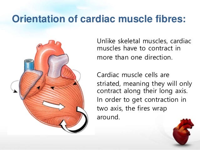
Cardiac Muscle Physiology
 www.slideshare.net
cardiac fibres skeletal
Cardiac Muscle (labeled) | Flickr - Photo Sharing!
 www.flickr.com
labeled
Class 12 Biology Questions ~ NEET Biology: Medical Entrance Biology
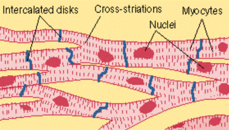 www.neetbiology.co.in
cardiac muscle tissue biology muscles cell diagram drawing cells questions answers class quiz multiple choice muscular neet entrance medical quizzes
Skeletal muscle histology tissue sarcomere cell file band bands sarcomeres myosin embryology animation showing light development isotropic unsw med edu. Cardiac muscles properties. Cardiac histology muscle tissues tissue lab slide cells human final photomicrographs anatomy quia physiology