external ear anatomy ct
CT Scan of the Temporal Bone: Overview, Normal Anatomy of the Middle. 16 Pics about CT Scan of the Temporal Bone: Overview, Normal Anatomy of the Middle : Yahoo, Congenital ear malformations and also Eustachian Tube – Oto Surgery Atlas.
CT Scan Of The Temporal Bone: Overview, Normal Anatomy Of The Middle
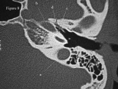 emedicine.medscape.com
temporal bone ct scan radiology anatomy ear assistant bulb axial inner normal jugular flashcards cram coronal middle carotid
Ear Anatomy
 www.edoctoronline.com
anatomy ear
Video/Image Link: Inner Ear
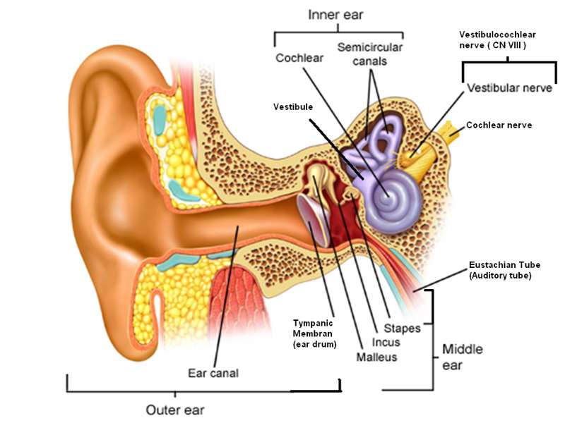 my.methodistcollege.edu
ear anatomy inner
File:Ear-anatomy.svg - Wikimedia Commons
 commons.wikimedia.org
anatomy ear svg file wikimedia commons 1300 1650 contribs talk kb
Yahoo
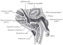 education.yahoo.com
anatomy ear external meatus auditory tube internal eustachian gray left right gland parotid bone section middle through opened wikidoc upper
Temporal Bone & Ear - Atlas Of Anatomy
 doctorlib.info
anatomy canal auditory ear external temporal bone semicircular plane atlas doctorlib medical info
Left Ear, 0° Endoscope: View Of The Prussak Space And The Surrounding
 www.researchgate.net
prussak endoscope endoscopic tympanic membrane malleus transcanal folds umbo tympani jove ligament malleolar chorda dissection discovering annulus
The Human Ear Extends Deep Into The Skull. Its Main Parts Are The Outer
 www.pinterest.com
ear inner anatomy middle human outer skull hearing parts speech body extends deep its into main health skulls references photons
The Radiology Assistant : Temporal Bone - Anatomy 2.0
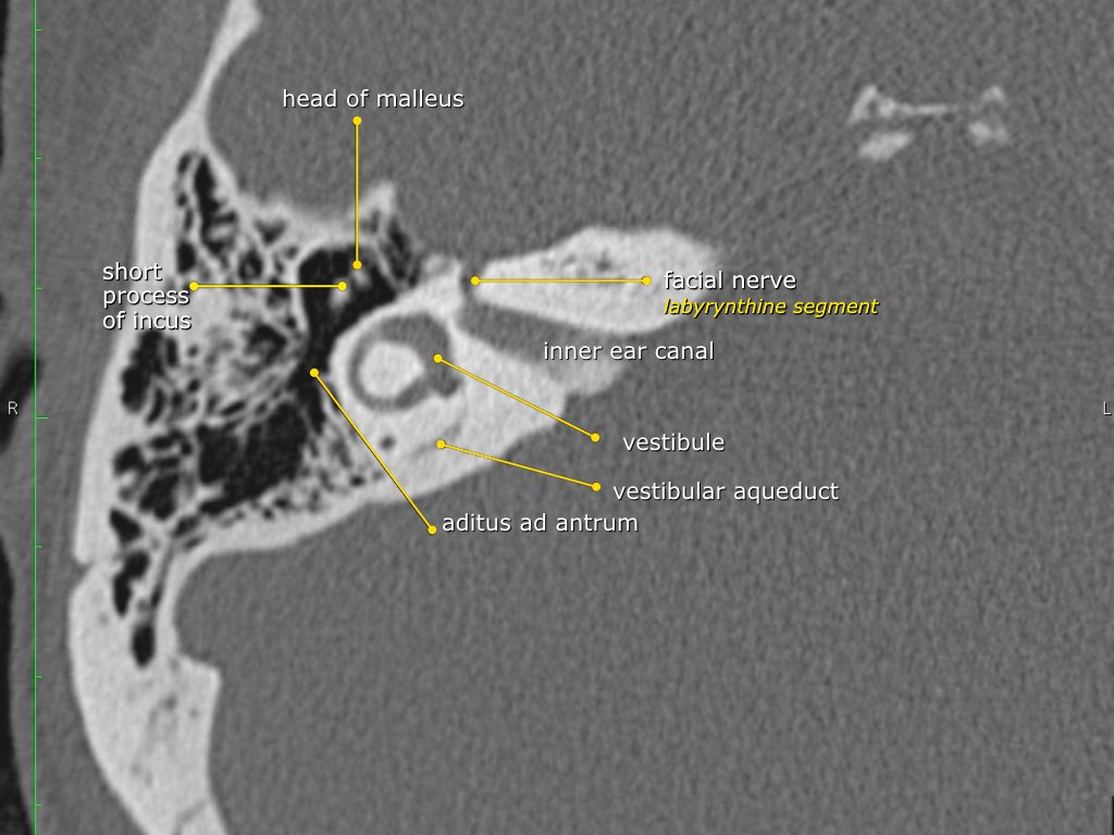 radiologyassistant.nl
anatomy bone temporal radiology assistant
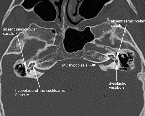
Congenital Ear Malformations
 uwmsk.org
ear charge syndrome atresia choanae retardation growth congenital axial abnormalities defects genital urinary deafness development
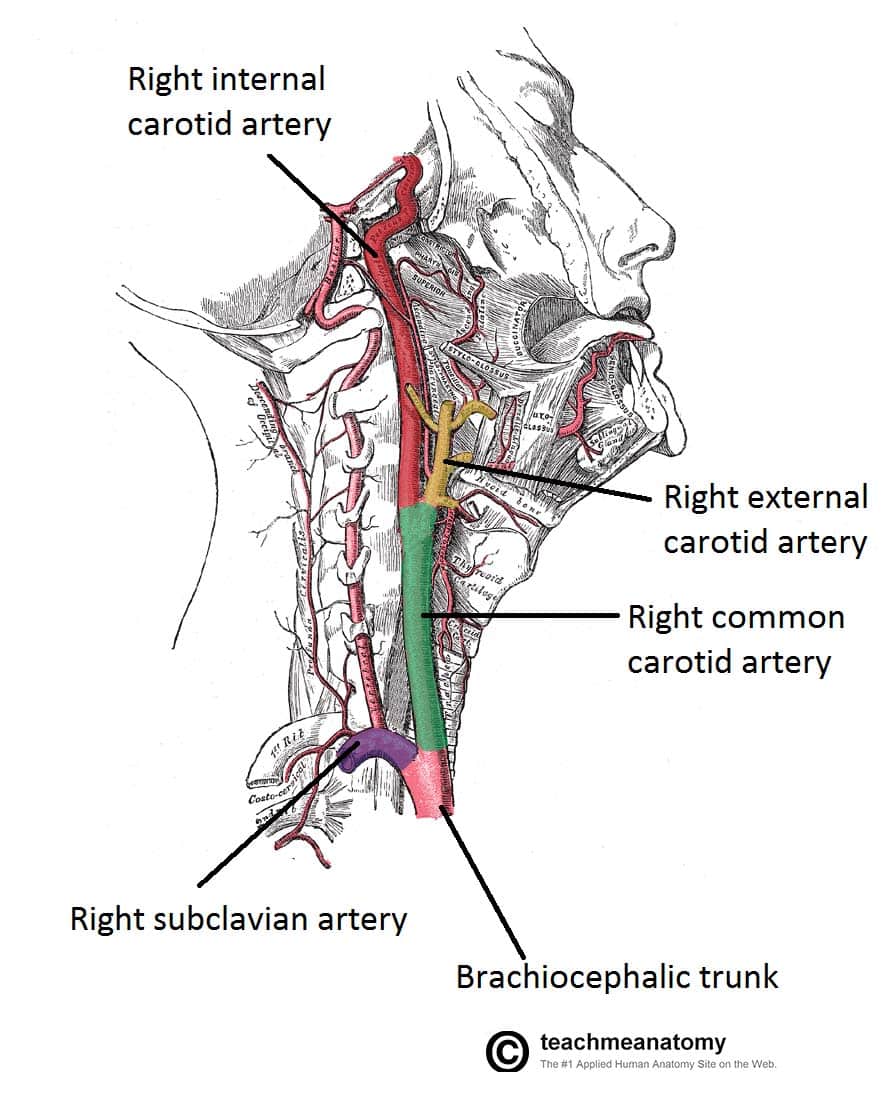
Blood Vessels And Lymphatics Of The Head And Neck - TeachMeAnatomy
 teachmeanatomy.info
carotid neck artery vessels arteries head diagram lateral blood common bifurcation veins vein teachmeanatomy anatomy major human showing branches left
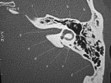
CT Scan Of The Temporal Bone: Overview, Normal Anatomy Of The Middle
 emedicine.medscape.com
temporal bone ct scan canal axial semicircular anatomy ear petrous radiology lateral normal cochlea auditory superior facial inner section assistant
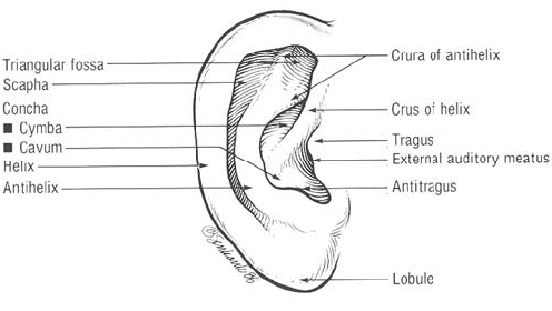
Medi Photos: Surface Anatomy Of The External Ear
 mediphotos.blogspot.com
ear external anatomy surface helix medi auricle auditory rim canal consists definition
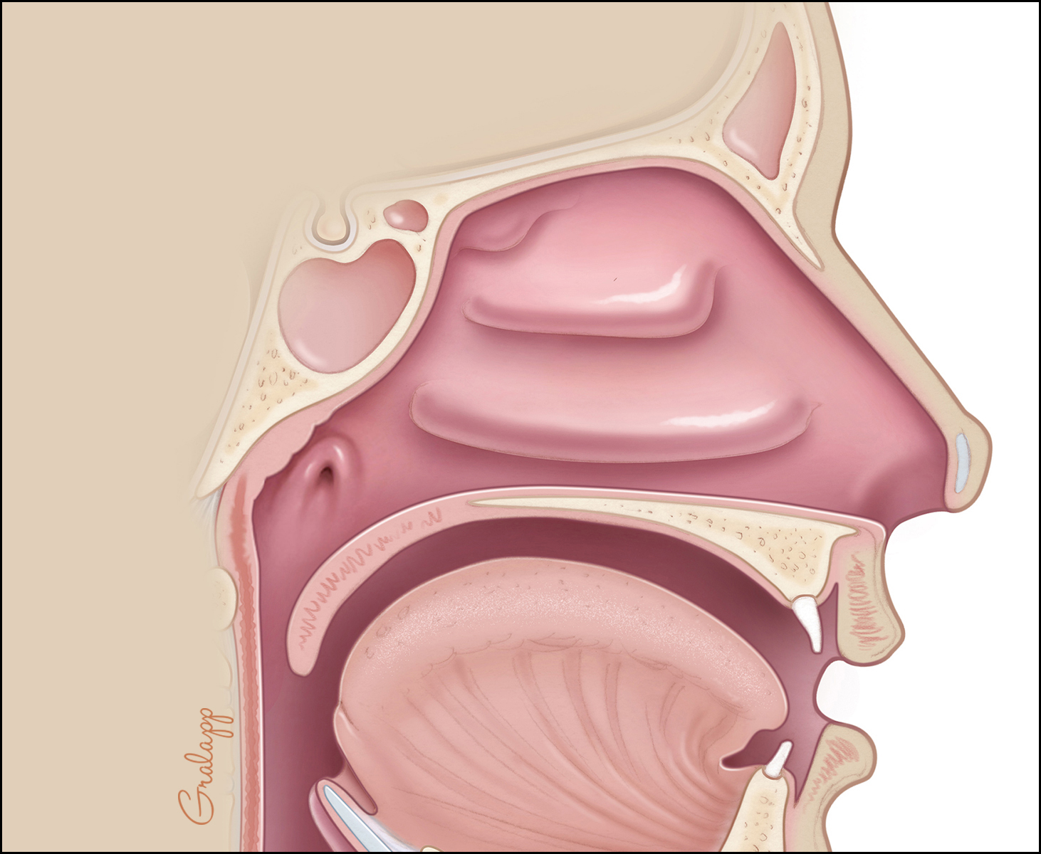
Eustachian Tube – Oto Surgery Atlas
 otosurgeryatlas.stanford.edu
tube eustachian anatomy ear orifice nasopharyngeal
Anatomy Of The Ear Flashcards - Cram.com
 www.cram.com
ear anatomy cram flashcards gland sublingual secretion submandibular
Viewing Playlist: Annotated Anatomy | Radiopaedia.org
 radiopaedia.org
annotated radiopaedia
Anatomy ear. Tube eustachian anatomy ear orifice nasopharyngeal. Viewing playlist: annotated anatomy