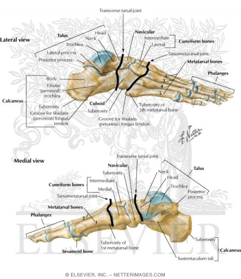
ankle bones diagram
lateral and medial view of the extrinsic muscles that move the foot and. 17 Images about lateral and medial view of the extrinsic muscles that move the foot and : left and right foot bones coloring pages, Bones of the foot and ankle, superior view with labels - Appendicular and also Learn all muscles with quizzes and labeled diagrams | Kenhub.
Lateral And Medial View Of The Extrinsic Muscles That Move The Foot And
 www.pinterest.com
muscles medial lateral extrinsic move foot muscle anatomy toes leg human body cf system muscular
Lower Limb And Pelvis | Radiology Key
 radiologykey.com
lower limb pelvis radiology fig radiologykey
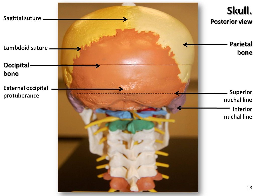
Multi-colored Skull, Posterior View With Labels - Axial Sk… | Flickr
 www.flickr.com
skull posterior skeleton axial labels atlas colored multi visual flickr english terms c1
Bones Of Ankle And Foot Unlabeled | Skeletal Anatomy | Pinterest | Bone
 www.pinterest.com
foot bones unlabeled ankle anatomy bone skeleton skeletal
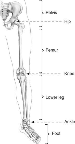
Blank Leg Bone Diagram / Skeletal System Labeled Diagrams Of The Human
 yeert-images.blogspot.com
sprain
Coronal Section Through The Ankle Joint | ClipArt ETC
 etc.usf.edu
ankle joint coronal section through etc clipart usf edu medium
Bones Of Ankle And Foot Unlabeled | PTA | Pinterest | Ankle, Anatomy
 www.pinterest.com
bones ankle foot anatomy unlabeled study human
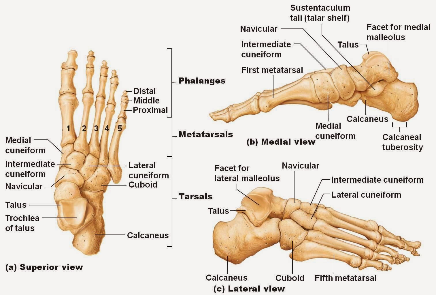
Bones Of The Foot And Ankle, Superior View With Labels - Appendicular
 flickr.com
foot bones ankle superior skeleton labels appendicular atlas visual english
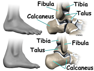
Ankle Bones
 cissink24.de
The Anatomy Of Ankle Bones - What You Should Know
 www.footvitals.com
Learn All Muscles With Quizzes And Labeled Diagrams | Kenhub
muscles diagram muscle learn labeled diagrams kenhub quizzes worksheets unlabeled worksheet anatomy unlabelled learning sponsored links
Tibia And Fibula | ClipArt ETC
 etc.usf.edu
tibia fibula clipart etc usf edu
A Patient’s Guide To Ankle Anatomy | Houston Methodist
 www.houstonmethodist.org
ankle anatomy talus foot leg called lower calcaneus dorsiflexion inside hinge bottom socket plantarflexion
AccessMedicine | Content | Ankle Anatomy, Anatomy, Human Body Anatomy
 www.pinterest.co.kr
foot anatomy ankle medial right leg fascia lower chapter body gross visit diagram med figure
Left And Right Foot Bones Coloring Pages
 freecoloringpages.co.uk
bones ankle diagram left foot right three joint coloring ankl
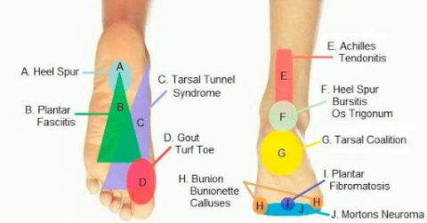
Taking A Look At Chronic Foot Pain | What Causes Foot Pain
 santarosapainandperformance.com
causes physiotherapy shrewsbury
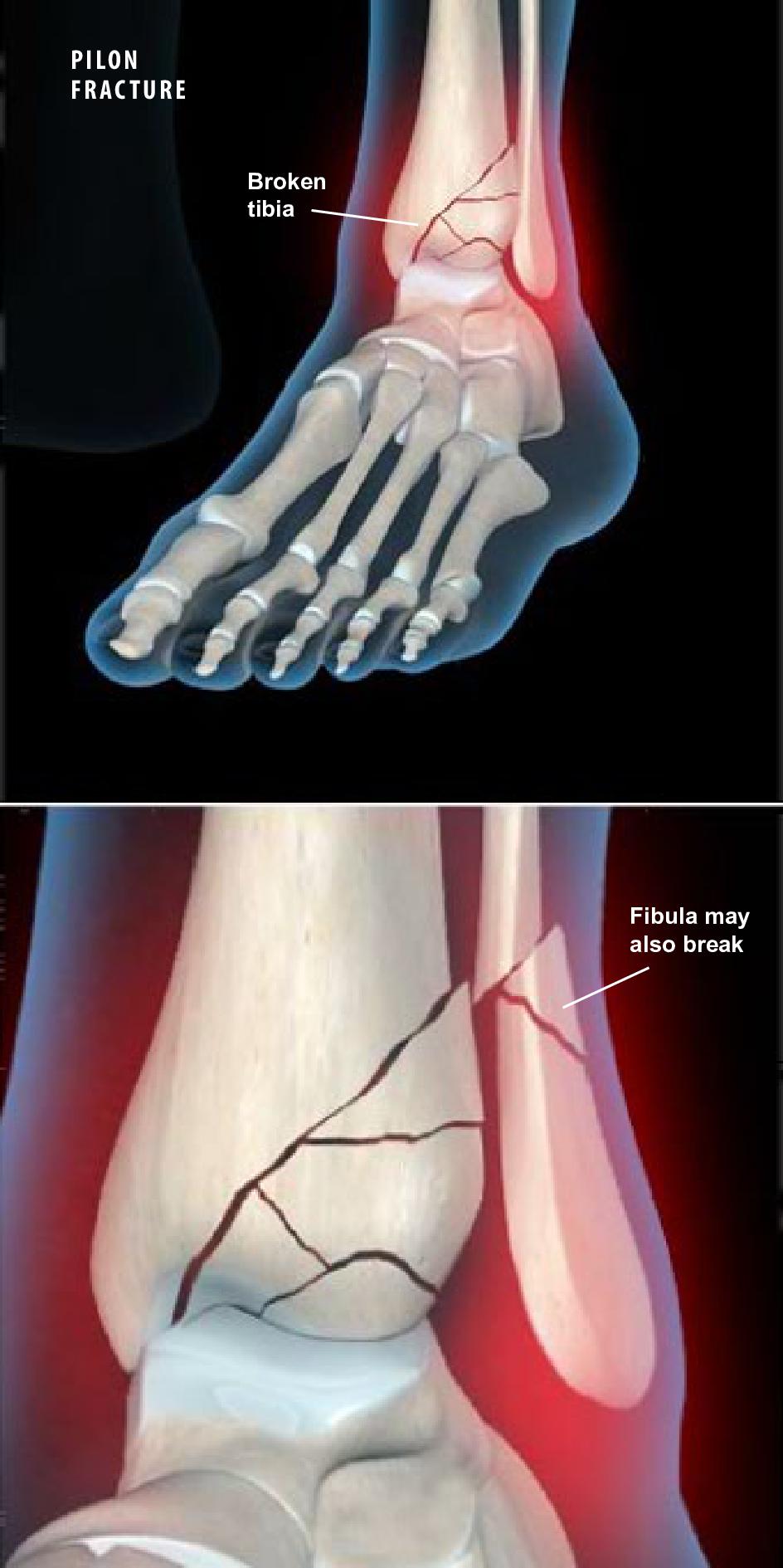
Pilon Fractures - Orthopaedic Associates Of Riverside
 www.orthoriverside.com
pilon fractures
Foot anatomy ankle medial right leg fascia lower chapter body gross visit diagram med figure. Taking a look at chronic foot pain. Bones ankle diagram left foot right three joint coloring ankl